

ImageProVision
Analytical Instruments
ImageProVision
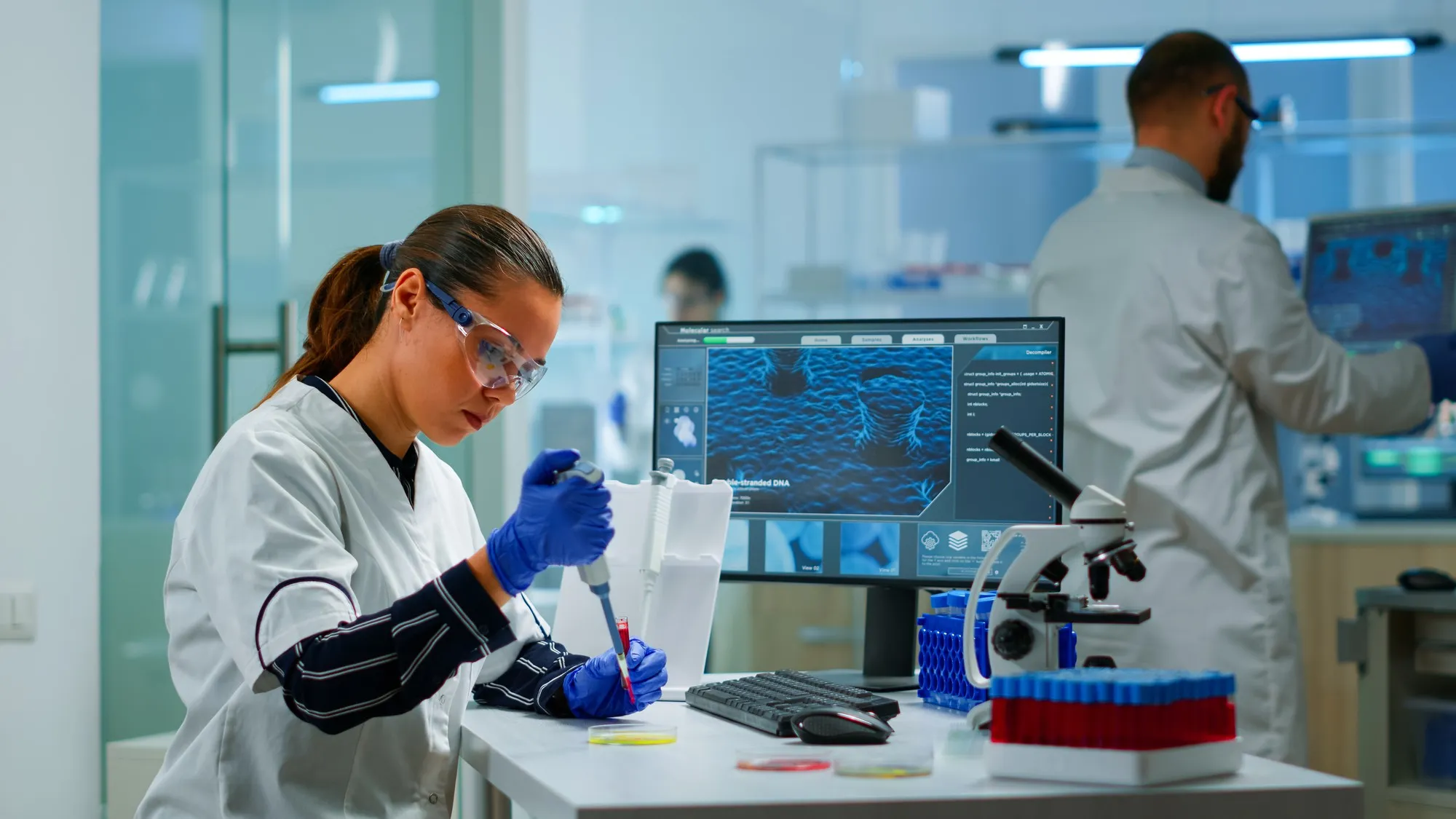
" The Next Level of Scientific Image Analysis " ImageProVision is a leading brand in high-precision image analytics and specialized in advanced processing that makes the microscopic world crystal clear, revealing details that were once impossible to see. This technology helps pharmaceutical companies, researchers, and manufacturers turn complex visual data into clear, actionable answers. By using ImageProVision, industries can speed up scientific discovery, stay compliant with regulations, and ensure every product meets the highest quality standards.
Product Gallery

Related Applications
API Particle Characterization
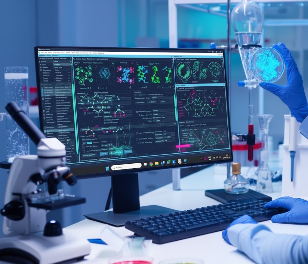
Precision Particle Morphology with ImageProVisionIn the pharmaceutical, food, and cosmetics industries, particle morphology—the structure and form of particles—is a critical quality parameter. Understanding shape and size is essential for identifying irregularities that can compromise the integrity of a final product.Advanced Analysis MethodsWhile traditional methods like light microscopy and SEM provide snapshots, ImageProVision leverages cutting-edge Image Analysis to deliver:High Throughput: Rapid measurement of thousands of individual particles.Superior Accuracy: Eliminating the human error associated with manual microscopy.Robust Insights: Reliable data across a diverse range of materials and formulations.The Impact on Pharmaceutical DevelopmentFor Active Pharmaceutical Ingredients (APIs), the particle-level details analyzed by ImageProVision are directly linked to how a drug performs. Characterizing properties like surface area, density, and refractive index allows for better prediction of performance and stability.How ImageProVision optimizes manufacturing:Flow & Uniformity: Ensures better powder flowability, blend uniformity, and compressibility.Enhanced Processing: Streamlines key steps such as granulation, mixing, and coating.Dissolution & Efficacy: Accurate Particle Size Distribution (PSD) ensures consistent drug solubility and therapeutic results.Overcoming the Agglomeration ChallengeA major hurdle in API analysis is agglomeration—where particles stick together and skew results. ImageProVision’s advanced image analysis software accurately distinguishes and measures individual particles within these clusters, providing a clear, dependable workflow where traditional methods fail.Regulatory Compliance & StandardsImageProVision solutions are designed to align with international regulatory standards, specifically supporting United States Pharmacopeia (USP) protocols:StandardMethodPrimary UseUSP <776>Optical MicroscopyAnalysis of size, shape, and crystallinityUSP <429>Laser DiffractionRapid measurement of particle size distribution
Excipient Particle Characterization
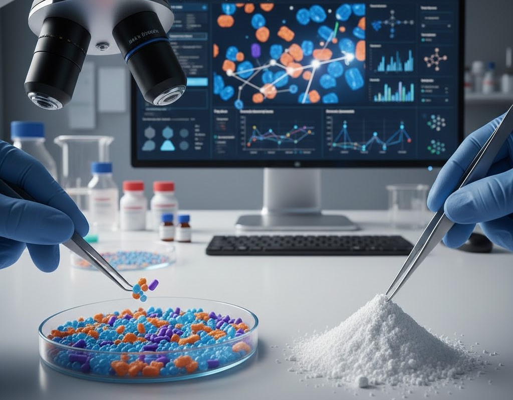
In pharmaceuticals, the “inactive” ingredients are just as important as the medicine itself. These substances, known as excipients, act as the binders, fillers, and diluents that hold a drug together.At ImageProVision, we help you analyze the physical and chemical properties of these particles to ensure your medicine works perfectly every time.Why Particle Analysis MattersThe size and shape of excipient particles determine how a drug performs. If particles aren’t “just right,” your formulation could face serious issues:Size Issues: Particles that are too large or too small can ruin the blending process and change how fast a drug dissolves in the body.Shape Issues: Irregularly shaped particles often lead to clumping, settling (sedimentation), or uneven mixing.Stability Risks: Poor particle quality can lead to “segregation,” where ingredients separate, or “poor wetting,” where the powder doesn’t mix well with liquids.The Benefits of Using ImageProVisionBy characterizing your excipients early with ImageProVision, you move away from trial-and-error and toward data-driven science.FeaturesBenefitsEarly DetectionSpot clumping or mixing issues before they ruin a batch.ConsistencyEnsure every batch of raw material from your suppliers meets the same high standards.Speed to MarketReduce the time spent on pre-formulation by picking the right materials the first time.Regulatory ReadinessBuild a solid database of “Functionality-Related Characteristics” (FRCs) for smoother regulatory submissions
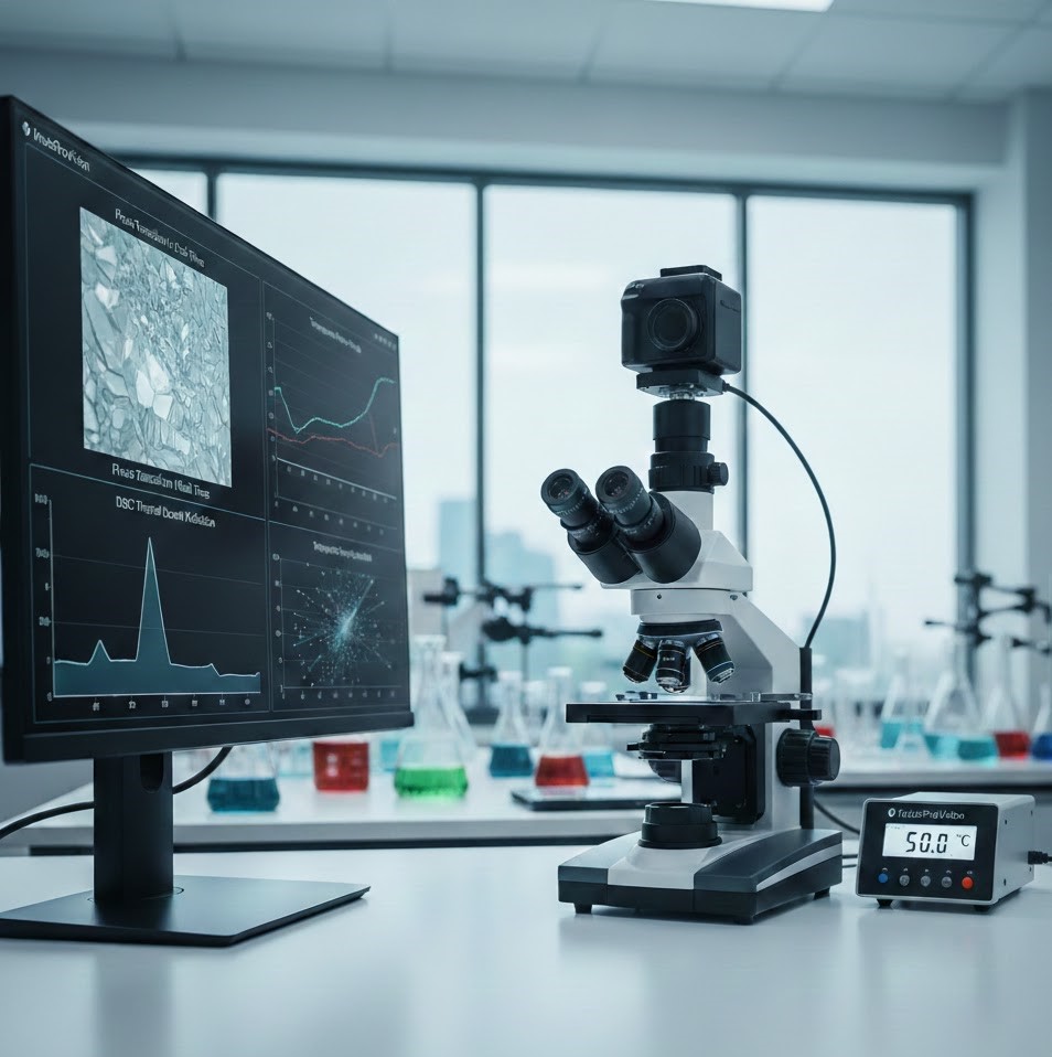
Hot Stage Microscopy
Hot-Stage Microscopy allows you to watch physical and chemical changes happen in real time. By combining controlled heating with high-powered visuals, the ImageProVision system gives you a front-row seat to the thermal life of your compounds.Why Use Hot-Stage Microscopy?While standard tests give you data points, Hot-Stage Microscopy gives you the “how” and “why.” It allows researchers to visually track:Phase Transitions: Watch exactly when a substance melts, solidifies, or changes form.Stability & Degradation: Identify the precise moment a compound begins to break down.Polymorphic Changes: Spot different crystal structures that could affect how a drug performs.Impurity Detection: See how contaminants interfere with a material’s thermal behavior.Critical for Pharmaceutical DevelopmentIn the world of drug formulation, temperature is everything. ImageProVision helps teams ensure product safety and efficacy by:Optimizing Formulations: Select the best ingredients (excipients) that stay stable during manufacturing and storage.Predicting Shelf Life: Understand how heat-sensitive compounds will behave over time.Checking Compatibility: Ensure that different components in a tablet won’t react negatively when exposed to heat.Reducing Delays: By spotting instability early, you can refine your formula before reaching expensive regulatory testing stages.The ImageProVision AdvantageImageProVision comprehensive system bridges the gap between raw data and visual proof. By integrating a precision hot-stage with advanced image analysis tools, ImageProVision enables you to:Identify Specific Particles: Easily find Active Pharmaceutical Ingredients (APIs) within complex mixtures or disintegrated tablets.Sync with DSC: Use our visual data to validate findings from Differential Scanning Calorimetry (DSC) for a complete thermal profile.Automated Analysis: Capture and analyze changes related to both temperature and time with professional-grade software
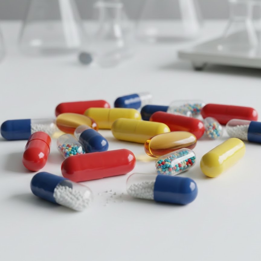
Capsule Seam Analysis
In softgel manufacturing, the “seam” is the line where the two halves of the capsule meet. If this seal isn’t perfect, the medicine inside could leak, spoil, or not dissolve correctly in the body.The ipvPSA system from ImageProVision automates the inspection of these seams to ensure every capsule is safe and effective.Why Seam Analysis MattersA capsule is only as good as its seal. If the seam is uneven, too thin, or misaligned, it can lead to several problems:Leakage: The liquid medicine inside can seep out.Durability: The capsule might break during packaging or shipping.Performance: A bad seal can change how fast the capsule dissolves, meaning the patient might not get the right dose at the right time.What the ipvPSA System ChecksManual inspection is a good start, but it’s prone to human error. The ipvPSA software uses high-tech imaging to measure specific details that the human eye might miss:MeasurementWhy it MattersSeam ThicknessEnsures the seal is strong enough to hold the liquid.Seam WidthChecks for consistency across the entire capsule.Seam AngleLooks for sharp or blunt edges that could cause mechanical failure.
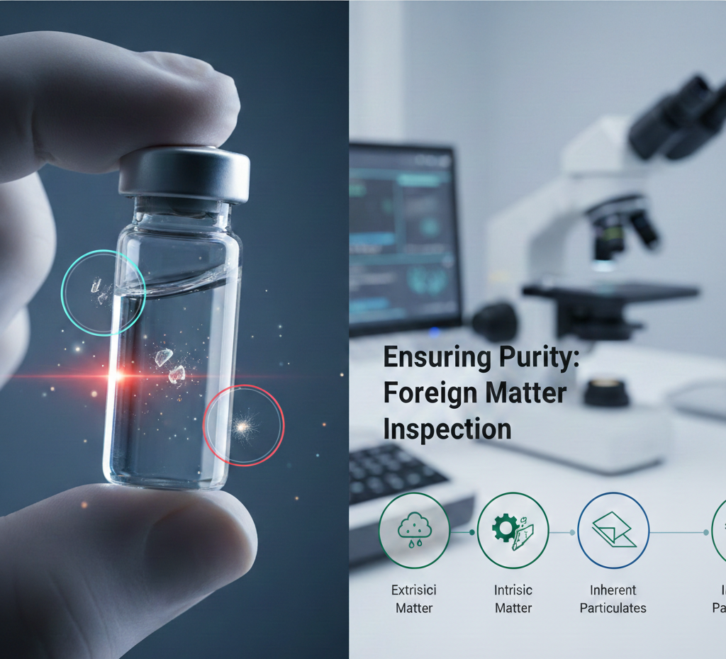
Foreign Matter Inspection (Vial Inspection - Intrinsic & Extrinsic)
In pharmaceutical manufacturing, even the smallest speck can be a major problem. ImageProVision specialize in the detection and classification of contaminants within vial-based products to ensure every dose is safe, stable, and compliant with global standards.Contaminants are generally categorized into three types. Identifying which one you’re dealing with is the first step in fixing the root cause:Extrinsic Matter: Particles from the outside world, such as dust, fibers, or environmental debris.Intrinsic Matter: Particles from the manufacturing process itself, like glass fragments, metal shavings, or leftover residues.Inherent Particulates: Matter originating from the packaging, such as vial delamination (flaking glass) or bits of rubber stoppers.How ImageProVision HelpsWe use a combination of advanced microscopy and automated tools to analyze the shape, size, and structure of particles. This “morphological analysis” allows us to pinpoint exactly when and where the contamination occurred.Our Inspection Process:Initial Screening: Using high-magnification light tools to scan for visible foreign matter.Isolation: Once a particle is found, it is isolated for closer study.Detailed Analysis: We evaluate optical properties and material composition to understand how the particle might interact with your drug formulation.Root Cause Support: Insights from our inspections help you refine your vial washing, rinsing, and siliconization processes.Why It MattersForeign matter isn’t just a cosmetic issue—it can ruin a drug’s effectiveness or cause dangerous reactions in patients. By following USP Chapter 1790 guidelines, ImageProVision helps you:Reduce Risks: Lower the chances of product recalls and regulatory delays.Maintain Integrity: Verify that your packaging and closure systems are working perfectly.Improve Quality: Use data-driven insights to choose better materials and improve your production line.Advanced Analytical TechniquesImageProVision employ industry-leading methods to give you total clarity:Light Obscuration (LO) for rapid counting.Membrane Microscopy for detailed visual confirmation.Automated Particulate Counting to eliminate human error.Compositional Analysis to identify leachables or manufacturing residues.
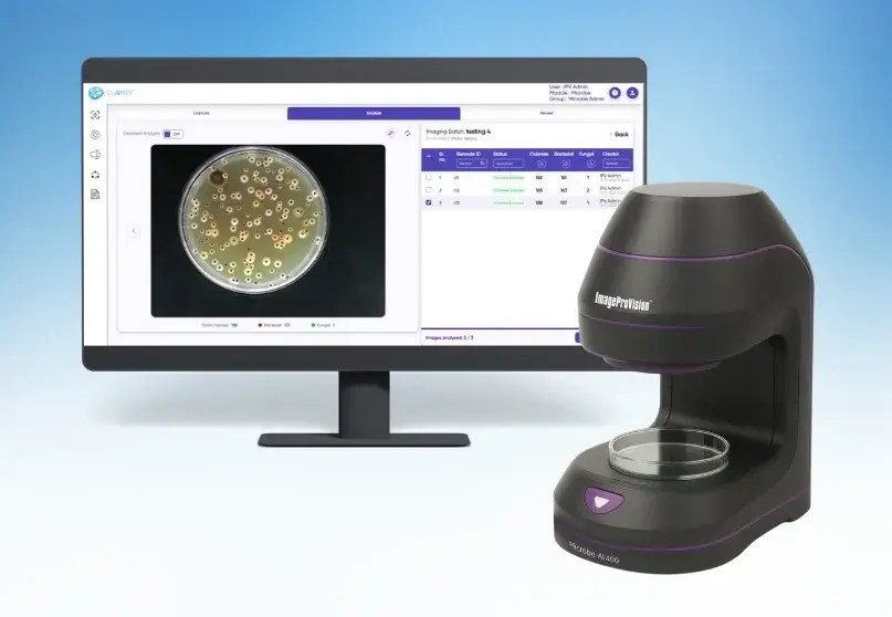
Microbiology Colony Counting and Gram Staining
ImageProVision specialize in providing the clarity and precision needed for advanced microbiological analysis.Whether you are working in a high-tech pharmaceutical lab or monitoring food safety, understanding the quantity and type of bacteria in your samples is vital. Here is a simple guide to how colony counting and Gram staining work together to safeguard your products.1. Colony Counting: Measuring the QuantityColony counting is the standard method for determining the “microbial load” or how much bacteria is present in a sample.How it works: A sample is spread onto a nutrient-rich plate. Each single, living bacterium grows into a visible cluster called a “colony.”The Goal: By counting these colonies, scientists calculate the concentration of bacteria in the original sample.The Challenge: Manual counting can be slow and prone to human error, especially with irregular or overlapping colonies.2. Gram Staining: Identifying the TypeWhile counting tells you how many, Gram staining tells you what kind. This process uses a specific series of dyes to categorize bacteria based on their cell wall structure.Gram-Positive (Purple): These bacteria have thick cell walls that retain the initial purple dye.Gram-Negative (Pink): These bacteria have thinner walls and take on a pink/red color from the final “counterstain.”Why it matters: This distinction is the first step in identifying a species and choosing the right antibiotic or disinfectant.Why Use Both? A Complete Microbial ProfileUsing these two methods together provides a full picture of your sample:Quantification: You know the scale of the growth.Classification: You know if you are dealing with a single type of contaminant or a complex mixture.Industry ApplicationsMicrobiology isn’t just for the lab; it’s essential for safety across multiple sectors:IndustryPrimary Use CasePharmaceuticalsValidating sterilization, testing drug efficacy, and meeting regulatory audits.Food SafetyMonitoring spoilage organisms and checking if preservatives are working.EnvironmentalTracking microbial levels in water, air, and soil to protect public health.Clinical ResearchMaking rapid decisions for infection control and tracking antibiotic resistance.Problems Addressed by ImageProVision Microbe AIThe “Microbe AI” platform is specifically engineered to solve inefficiencies in traditional laboratory workflows:Manual Counting Errors: Traditional “eye-balling” of Petri dishes is prone to human error and fatigue. The AI provides high-precision CFU (Colony Forming Unit) countinTime Consumption: It automates the analysis of microbial growth, significantly reducing the time required for sample processing.Complex Morphology: The AI can distinguish between different types of microbial colonies, handling overlaps and varying sizes that are difficult for basic software to separate.Data Integrity & Compliance: It generates digital records and audit trails, helping pharmaceutical labs meet 21 CFR Part 11 and other regulatory requirements.
Deformulation Analysis
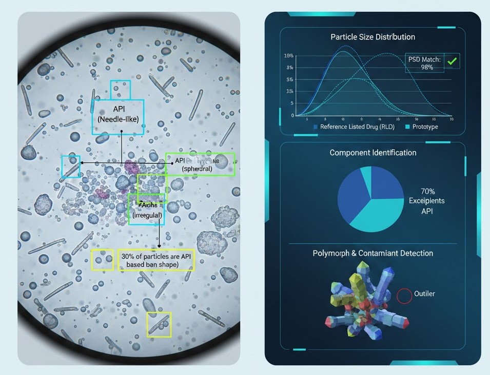
Deformulation, or Reverse Engineering, is the process of breaking down a product’s formulation to identify and quantify its components (APIs and Excipients). ImageProVision (IPV) transforms this traditionally manual, chemistry-heavy process into a faster, data-driven workflow using AI-powered microscopy and image analytics.Here is how their technology addresses the core challenges of deformulation: Particle Morphological FingerprintingThe foundation of deformulation is identifying which particle is which. IPV’s software uses Morphologically Directed Analysis to categorize particles based on their physical “fingerprints.”The Problem: In a blended tablet, the API (Active Pharmaceutical Ingredient) and excipients are mixed. Traditional methods struggle to tell them apart visually.The IPV Solution: By analyzing parameters like circularity, aspect ratio, and convexity, the AI creates a library of shapes. It can then scan a sample and say, “30% of these particles are API based on their unique needle-like shape.”Component Identification & QuantificationThrough products like ipvAutoM, the system automates the counting and measurement of individual components within a complex mixture.Solid State Analysis: It helps determine if the API is in a specific crystalline or amorphous state, which is critical for bioequivalence.Volume Distribution: Beyond just counting, it calculates the volume and mass distribution of components, allowing formulators to replicate the exact “recipe” of a reference listed drug (RLD).Particle Size Distribution (PSD) MatchingMatching the PSD of the original product is vital for ensuring the generic version dissolves at the same rate.FeatureHow ImageProVision HelpsAgglomerate DetectionAI distinguishes between a single large particle and a “clump” of smaller ones, preventing skewed data.Automated ScanningRapidly scans thousands of particles to provide a statistically significant representation of the RLD.Comparison ToolsOverlays the PSD of your prototype against the RLD to identify exactly where the formulation needs adjustment.Polymorph & Contaminant DetectionDuring deformulation, you must identify different polymorphic forms of an API, as these affect stability and solubility.AI Classification: The software can be trained to recognize the visual texture and boundaries of specific polymorphs that are otherwise invisible to the naked eye or hard to distinguish via standard XRD (X-Ray Diffraction) alone.Foreign Particle Identification: It quickly spots impurities or “outlier” particles that don’t belong in the standard formulation profile.Summary of BenefitsSpeed: Reduces deformulation timelines from weeks to days.Accuracy: Eliminates human bias in particle characterization.Regulatory Compliance: Provides the digital “proof” and high-resolution images required by regulatory bodies (like the FDA) to prove bioequivalence.
Globule Size Determination in semi-solid dosage forms
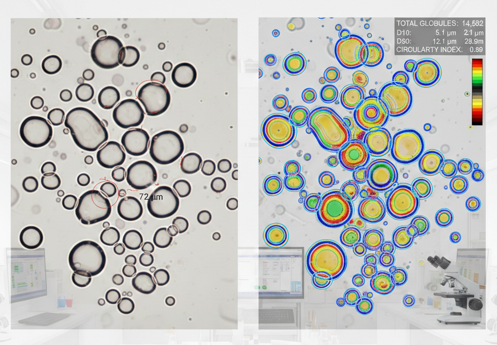
Globule size determination is a critical quality attribute (CQA) for semi-solid dosage forms like creams, ointments, and emulsions. If the oil droplets (globules) are too large or inconsistent, the drug may not be absorbed correctly through the skin, or the product may become unstable and separate.ImageProVision automates this complex analysis through its AI-driven microscopy software, specifically designed to handle the “messy” visual nature of semi-solids.How ImageProVision Automates Globule AnalysisAutomated Phase SeparationIn a cream or emulsion, globules often overlap or “cluster” together. Standard software often miscounts these clusters as one giant globule.AI Edge Detection: ImageProVision uses deep learning to identify the precise boundaries (the “edges”) of each individual globule, even if they are touching.Feature Extraction: It distinguishes between the continuous phase (the base) and the dispersed phase (the globules) with high contrast enhancement.Statistical Accuracy (D10, D50, D90 )For regulatory filings (like ANDAs for generics), you must provide a statistical profile of the globule population.Massive Sample Size: Instead of a lab technician manually measuring 100 globules with a reticle, the AI can scan and measure thousands of globules in seconds.Automatic Calculation: It immediately generates the cumulative distribution:D50 :The median diameter (50% of globules are smaller than this value).D90 :Indicates that 90% of the distribution is below this size, ensuring there are no “large” outliers that could cause grittiness.Handling Non-Spherical & Deformed GlobulesSemi-solids are highly viscous, which can cause globules to appear non-spherical or elongated.Shape Factors: The software calculates “Circularity” and “Equivalent Circular Diameter.” This ensures that even if a globule is slightly squashed, its true volume is accurately represented.Comparison: Manual vs. ImageProVision AIFeatureManual MicroscopyImageProVision (ipvAutoM)Speed1–2 hours per slide< 2 minutes per slideBiasHigh (Human selects “easy” globules)Zero (Analyzes every visible globule)ReproducibilityLow (Varies between analysts)High (Standardized AI algorithm)Data IntegrityManual logbooks (Risk of error)Electronic records (21 CFR Part 11)Addressing Regulatory Requirements (FDA/EMA)Regulators require proof that a generic semi-solid is “bioequivalent” to the brand name. ImageProVision provides the raw image data and morphological reports necessary to prove that your formulation has the same internal structure and globule distribution as the Reference Listed Drug (RLD).

Particulate Matter Count - Filter Paper Analysis
Particulate Matter (PM) analysis on filter papers is a critical safety test for injectables, infusions, and medical devices. Traditionally, this is done by a technician staring through a microscope for hours, which is both exhausting and prone to “counting fatigue.”ImageProVision (specifically through ipvAutoM ) automates this by turning the filter paper into a digital map. Here is how the AI handles the heavy lifting:Full-Filter Scanning & StitchingA standard microscope view only shows a tiny fraction of a 47mm or 25mm filter paper.Automated Stage Control: The software moves the microscope stage in a “snake” pattern to capture every square millimeter of the filter.Image Stitching: It seamlessly joins hundreds of individual photos into one high-resolution “macro” image of the entire filter. This ensures no particle is counted twice and none are missed at the edges of a frame.Particle Detection & ClassificationThe AI doesn’t just “see” a dot; it analyzes the morphology to categorize the debris.Size Binning: It automatically sorts particles into the standard regulatory bins.Fiber vs. Particle: The system uses “Aspect Ratio” and “Length” to distinguish between a round speck of dust and a long lint fiber. This is crucial because fibers often have different regulatory limits or source origins.Color & Transparency: It can detect transparent “proteinaceous” particles or colored plastic shards that might be invisible to basic thresholding software.Advanced Hardware IntegrationTo get the best data from a filter paper, ImageProVision uses specific lighting techniques:Polarized Light: Helps identify birefringent materials (like certain plastics or crystals).Top/Episcopic Illumination: Essential for opaque filter papers to ensure the particles “pop” against the background.Meeting Regulatory Standards (USP/EP/JP)The software is pre-configured to generate reports that satisfy global pharmacopeia standards:StandardFocus AreaImageProVision RoleUSP <788>Particulate Matter in InjectionsAutomated counting/sizing for sub-visible particles.USP <790>Visible ParticulatesValidation of manual inspection via high-res imaging.21 CFR Part 11Electronic RecordsProvides encrypted audit trails, e-signatures, and raw image storage.Summary of Key BenefitsEliminates Human Bias: Two different technicians often give two different counts; the AI provides a reproducible, objective number.Archivability: Instead of throwing the filter paper away or watching it degrade, you have a permanent digital twin of the test for future audits.Speed: What takes a human 30–45 minutes, the AI completes in under 5 minutes.